
Translational Bioimaging Resource (TBIR)

Bioscience Research Laboratory
1230 N Cherry Ave.
Tucson AZ, 85719
TBIR serves as a university-wide resource for pre-clinical biomedical imaging. The resource has the capability to image small biological constructs, small and large animals, and humans. For imaging small biological constructs and small animals the TBIR includes intra-vital microscopy, MRI, ultrasound, CT, PET, SPECT, and bioluminescence. For imaging larger animals and humans the resource includes MRI and ultrasound. The resource includes personnel to help you with project development and to help you use the equipment. Please contact TBIR@arizona.edu to learn more about TBIR's imaging resources and services.
Visit this link for facility access : https://bbic.arizona.edu/resources/mri-facility-access
Human MRI Facility
The MRI facility is part of the Translational Bioimaging Resource (TBIR) and is housed in the basement of the Biosciences Research Labs (BSRL) building. There is currently one MRI machine available with two more machines that will be available for use later in 2025.
Siemens Skyra 3T MRI Machine
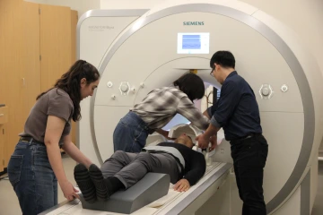
The Siemens Skyra 3T has Syngo MR VE11c software (as of Nov 14, 2018). 7 new licenses have been installed (3D ASL, Abd Dot, Freeze IT, Liver Lab, Multiband, MyoMaps, Resolve, SMS). In addition, several WIPs (beta sequences) have been updated to work with the new VE11C software. A 32 channel head coil and a 20 channel head coil are available.
Please treat all the equipment carefully! This equipment is a shared resource that is used daily (and sometimes nightly) by multiple labs. The equipment is highly specialized and, therefore, very expensive, and many components and cables are fragile.
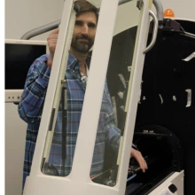
Take the MRI Scanner Virtual Tour
Siemens Cima.X 3T MRI Machine
A new Siemens 3T scanner was installed in March 2025 and will be available for use later this year. We will have updates on this state-of-the-art machine coming soon.
Siemens Free.Max .55T MRI Machine
A lower strength 0.55T scanner will be installed in the summer of 2025 and will be available for use in the fall.
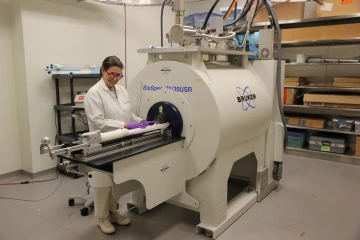
Small Animal Imaging
The TBIR small animal imaging facility supports the research community at the University of Arizona and industry partners in pharmaceuticals, health care, veterinary care, and behavioral sciences. TBIR regularly works with researchers in the UA’s College of Science and College of Medicine, among others, as well as industry partners in pharmaceuticals, health care, veterinary care, and behavioral sciences.
TBIR supports research that addresses a wide range of research questions, including questions related to arthritis, traumatic brain injuries, PTSD, cancer treatment efficacy, depression, kidney transplant viability, CPR, Alzheimer’s, complex grief, and healthy aging.
The Lab manages a 7T Bruker Biospec research MRI scanner and a newly established PET/MRI capability. With these modalities, users can perform physiological and functional imaging of soft tissue anatomy, tumor growth, angiogenesis, cellularity, inflammation, myelination, myocardial function, early therapeutic effects, extracellular pH, redox state, and other applications. 3D anatomical imaging of soft tissues is performed at up to ~300 μm resolution. The facility has technical support to aid in surgical procedures and anesthesia, protocol modifications and compliance, safety training, and consultations.
The PET/MRI insert by Cubresa, Inc. enables evaluation of tumor therapy response through diffusion-weighted MRI with PET tracers specific for tumor proliferation, improved tumor detection through diffusion-weighted and T2-weighted MRI with PET tracers for specific biomarkers, evaluations of drug delivery and perfusion for molecular theranostics evaluations.
The Lab regularly works with researchers in the University of Arizona’s College of Science, College of Medicine, and College of Pharmacy, as well as industry partners dedicated to research and development in biomedicine and oncology.
High-Resolution Ultrasound Imaging
HRUF provides small-animal ultrasound imaging with axial resolution down to 30 microns. With the VisualSonics Vevo 2100, as well as data management and analysis software, the system can be used for biomedical research and material characterization.
The HRUF regularly works with researchers in the UA’s College of Medicine, Arizona Cancer Center, Sarver Heart Center, College of Optical Sciences, and College of Engineering, as well as a variety of industry partners, that participate in biomedical research and material characterization.