Please feel free to use the following language to add to your imaging grants:
Facilities:
Translational BioImaging Resource (TBIR)

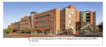
The Translational Bioimaging Resource (TBIR) is housed in the basement of the BSRL building. The 12,000 sq. ft. TBIR serves as a university-wide resource for pre-clinical biomedical imaging. The resource has the capability to image small biological constructs, small and large animals, and humans. For imaging small biological constructs and small animals, the TBIR includes intra vital microscopy, MRI, ultrasound, CT, PET, SPECT, and bioluminescence. For imaging larger animals and human the resource includes MRI and ultrasound. The resource includes personnel to help researchers with project development and to help use the equipment. Below are some of the major resource provided by the TBIR:
- 3T MRI scanner for human and large animals: The TBIR is dedicated to supporting MRI research within the University community and for industry partners in pharmaceuticals, health care, veterinary care, and behavioral sciences. The TBIR houses a 3 Tesla Siemens Skyra VE11C that is able to support work that addresses a wide range of research questions, including questions related to arthritis, traumatic brain injuries, PTSD, cancer treatment efficacy, depression, kidney transplant viability, CPR, Alzheimer’s, complex grief, and healthy aging. Dr. Chen has installed Pulseq environment in this 3 T MRI system, so that the MRI-stim pulse sequences (in the Pulseq format) can be executed directly in this MRI system.
- Small Animal MRI: The TBIR manages a 7T Bruker Biospec research MRI scanner. Users can perform physiological and functional imaging of soft tissue anatomy, tumor growth, angiogenesis, cellularity, inflammation, myelination, myocardial function, early therapeutic effects, extracellular pH, redox state, and other applications. 3D anatomical imaging of soft tissues is performed at up to 300 μm resolution. The facility has technical support to aid in surgical procedures and anesthesia, protocol modifications and compliance, safety training, and consultations.
- A Cubresa NuPET MRI compatible PET insert enables evaluation of tumor therapy response through diffusion-weighted MRI with PET tracers specific for tumor proliferation. The PET insert can also improve tumor detection through diffusion-weighted and T2-weighted MRI with PET tracers for specific biomarkers as well as evaluate drug delivery and perfusion for molecular theranostics assessments.
- High Resolution Ultrasound: The TBIR manages a VisualSonics Vevo 3100 along with data management and analysis software. This small-animal ultrasound device provides axial resolution down to 30 microns. The system can be used to measure a variety of cancer-related metrics such as tumor growth, tumor blood flow and volume, angiogenesis, and molecular imaging with microbubble contrast agents. Cardiovascular imaging and measurement of cardiac function are two other popular applications. The technology also applies to other disciplines including neurosciences, embryology, and ophthalmology. Specialized human studies are also possible with institutional review board approval.
- MicroCT: Through a partnership with the University of Arizona Cancer Center, the TBIR provides a Siemens Inveon micro-CT scanner, which is a variable zoom cone-beam X-ray CT system that can generate images with a spatial resolution as small as 20 microns over a field of view as large as 8.4 cm x 5.5 cm. The large field of view enables single-bed position imaging of an entire mouse as well as larger objects such as rats with bed-stitching. The fully shielded design has built-in anesthesia and optional physiological monitoring. The scanner is best at evaluating bone density, but it can also image soft tissues with the use of CT contrast agents. Physiological monitoring ensures viability of the animal during the scan, and respiratory & cardiac gating improves image quality of the heart and lungs.
- Bioluminescence and Fluorescence Imaging: Through a partnership with the University of Arizona Cancer Center, the TBIR provides a Spectral Instruments Imaging Lago, which has powerful multi-modality imaging capabilities in an advanced, user-friendly system. The turn-key system offers bioluminescence and fluorescence imaging. The high performance 2048x2048 air-cooled CCD camera provides high sensitivity with low background noise for accurate quantitative analysis. The Lago has 14 excitation wavelengths ranging from 360-805 nm and 20 emission filters ranging from 490-870 nm. The system has a field-of-view ranging from 6x6 cm up to 25x25 cm. These dynamic filter combinations can support a wide range of applications ranging from well-plate imaging to small animal imaging. Bioluminescence imaging of reporter genes such as luciferase and green fluorescent protein are easily accomplished with this scanner, and imaging of exogenous fluorescent agents can also be performed. The Lago incorporates a heated imaging platform and built-in isoflurane gas anesthesia.
- Quantitative Digital Histology: Through a partnership with the University of Arizona Cancer Center the TBIR offers access to Definiens Tissue Studio 4.2 for biomarker and morphological profiling in histological images. Tissue Studio is an easy to use, robust, and powerful tool to automatically detect regions of interest based on user-defined criteria. It can distinguish cells and sub-cellular objects within target regions and determine morphology and expression profiles per individual cell or cell compartment. It is compatible with brightfield or immunofluorescence images and whole tissue slides or tissue microarrays. Tissue Studio is compatible with most major file formats including Aperio (.svs), Hamamatsu (.ndpi), Leica (.scn), Roche Ventana (.bif), Zeiss (.czi), and TIFF.
Computer Resources
In addition to dedicated lab computers, UA labs have essentially unlimited access to the high performance computer (HPC) systems hosted by the University of Arizona’s Research Data Center – a 1200 ft2 raised floor data center designed for water-cooled racks dedicated to centrally managed research computing systems and large grant funded systems, with capacity for 40 standard racks. RDC capacity allows UITS to maintain two generations of centralized large computer clusters simultaneously, providing continuity of research while each new system is phased in. Cooling in the RDC is both in-rack cooling with chilled water heat exchangers and 70 tons of Computer Room Air Conditioning (CRAC) units.
The HPC consulting staff has expertise that includes; support for scaling issues, debugging, parallelizing code, optimizing code to hardware, code clinics and new user workshops. HPC support staff handles the maintenance of the clusters including; software upgrades, security patching, hardware service, community software installation and licensing support.
UA provides centrally managed and maintained high performance research computing systems to all campus faculty and researchers. The following systems are available at no cost to researchers, and can provide high priority to researchers who have purchased dedicated nodes in the HPC.
Ocelote, a general-purpose cluster (purchased 2016) with 10000 cores, 71TB of Memory, FDR Infiniband (IB) Interconnect, 10G ethernet. Additionally, provisioned with a 336 core, 2TB virtual shared memory (vSMP) node and a 48 core, 2TB RAM dedicated node.
ElGato is a specialized Nvidia GPU high-performance cluster purchased with an NSF MRI grant awarded to faculty in Astronomy and the School of Information: Sciences, Technology, and Arts (SISTA.) The MRI grant stipulates that 30% of system resources be available for general campus research. This configuration includes 2176 Intel cores, FDR IB interconnect, 26TB of RAM, 140 Nvidia Tesla K20X GPUs, and 40 5110P Intel PHIs.
BIO5 Institute
The Bio5 institute supports interdisciplinary collaboration through pilot grants and shared equipment grants. Students and trainees in the lab benefit from the strength of all these affiliations and from working side-by-side with two graduate students from departments of Applied Mathematics, Biomedical Engineering, and Computer Engineering.
Rodent Phenotyping Core:
This new core, created for heart, vascular and lung research, provides cutting-edge resources and expertise to investigate genetically altered mice that mimic human disease. The core offers investigators full service support of in vivo and in vitro physiology studies in normal, exercise and diseased states through surgical models (MI, TAC, ACF), imaging techniques, and functional physiological assessments. The Core utilizes state-of-the-art facilities and equipment. Some examples include: microsurgical operating room, Visualsonics Vevo 2100 echocardiography system, Scisense pressure-volume admittance catheters for cardiac catheterization and pressure-volume analysis, DSI Telemetry (heart rate, ECG, blood pressure), MRI, whole body plethysmography for non-terminal respiratory studies, FlexiVent System apparatus for invasive measurements of respiratory mechanics and lung function, ex-vivo and in-vitro muscle mechanics and single cells physiology. The Director of this core is Dr. Henk Granzier and is managed by a full-time PhD scientist, Dr. Joshua Strom (who expertly performs and/or trains others in all procedures).
Center for Innovation in Brain Science at the UA Health Sciences
The Center for Innovation in Brain Science will accelerate the advancement of evidence-based clinical care of brain disorders caused by disease, genetics or trauma. The Center will focus on research and scholarship across the spectrum of brain disorders and the emerging area of brain and cognitive development, with particular emphasis on four areas of current UAHS strength: cognitive aging in health and disease, chronic pain and traumatic brain injury, stroke and aphasia, and integrative/systems neuroscience. The Center, led by Dr. Roberta Brinton, will speed the development of novel, multidisciplinary approaches to address neurodegenerative diseases through research, clinical practice interventions, education, and community collaborations.
Evelyn F. McKnight Brain Institute
This Center was founded in 2006 and is one of only four McKnight Institutes nationally. Researchers in this Center to understand normal changes in the brain as it ages, in the hopes of developing practical lifestyle recommendations and treatments that will lead to better memory, and longer, fuller cognitive lives for all. To do this, scientists are engaged in high-level neuroscience through the continual development of new technologies to better understand the brain in health and disease.
Bioinformatics
The Informatics/Bioinformatics Shared Service at The UA supports research in the following areas: genomics analysis (e.g. next generation sequencing, RNA-Seq, Chip-Seq and gene expression arrays, comparative genomic hybridization, DNA methylation, RNAi screens, genome and sequence analysis), genetic studies (SNP analysis and exome sequencing), proteomics, and other types of molecular data sets for cells and tissues, biological interpretation of the above data, including network, pathway and ontology analysis, systems analysis, genetic vulnerabilities for drug targeting, predictive patterns for outcome and data modeling, informatics support in the form of tissue and molecular databases, genome databases, and data sharing tools, and data integration of clinical, molecular, and genetic data utilizing CaBIG tools.
Center for Biomedical Informatics and Biostatistics (CB2):
CB2 has a dual mission of promoting research and offering services in the fields of biomedical informatics and biostatistics. Senior UA professors of Biomedical Informatics and Biostatistics currently form an internal advisory board promoting the active recruitment of tenure track faculty members in these quantitative sciences. Altogether, more than 20 staff biostatisticians, bioinformaticians, and biomedical informaticians work synergistically to conduct biostatistical studies, epidemiological analyses and research design as well as biomedical informatics analyses that comprise data from expression arrays analyses, high-throughput sequencing, proteomics, clinical warehouse, tissue specimen management systems, clinical trial management systems, and other biomedical data sources. CB2 is located in approximately ~3000 sq. ft. in the BIO5 Institute (in Keating building, where Dr. Chen’s office is located) and in the Cancer Center of The University of Arizona with access to shared facilities including 18 conference rooms with projectors. All members of CB2 have modern computing workstations with access to software bundles (R, SAS, STATA, Python, MS Visual Studio, REDCap, among others).
The University of Arizona Health Sciences (UAHS) Tucson, Arizona
University of Arizona Health Sciences (UAHS), is an educational, research, and patient-care network including: 5 University of Arizona Colleges (Medicine – Tucson, Medicine – Phoenix, Nursing, Pharmacy, and Public Health); 3 medical centers; and 23 centers and institutes dedicated to research and patient care. The UAHS Office of Research Administration provides concierge-level service to all UAHS investigators in support of research pre- and post-award activities, including customized grant searches for investigators using PIVOT, Grants.gov, Community of Science, REDCap and other sources, sponsored project application submission, contract negotiation, clinical trials, and compliance support in collaboration with Sponsored Projects Services, Office of Contracting & Research Services, and the IRB.
The University of Arizona
The University of Arizona (UA) is the leading public research university in the American Southwest and an ideal transdisciplinary research community for the proposed work. UA produces more than $580 million in annual research and ranks 20th among public universities according to the National Science Foundation. Enrolling more than 42,000 students representing every state in America and over 100 other countries in over 350-degree fields, this is a diverse community of people who thrive on innovation and collaboration between students, faculty and researchers from many diverse disciplines. The 387-acre Tucson campus includes 184 buildings: 159 on the main campus and 25 buildings in the adjacent Health Sciences Center complex.
Equipment
Magnetic Resonance Imaging system
The proposed research will be performed with a research-dedicated 3 Tesla Siemens Magnetom Skyra whole-body MRI system running VE11c software. The scanner features a short (163cm) and wide (70cm ID) magnet bore, a Direct RF Transmit/Receive amplifier rated to 37.5 kW, and a TIM [204x48] XQ gradient deck capable of 200T/m/s per axis slew rate with a minimal rise time of 225 microseconds and a maximum strength of 45 mT/m. The Skyra has the NeuroSuite software, which includes the fMRI/DTI package, allowing online processing and post processing of statistical maps, superimposing maps on cut planes through 3D volumes, data quality monitoring based on B0 field maps, tractography, FA, ADC, ASL perfusion weighting and CBF, SWI, 2D Spectroscopy, and 3D CSI. Coils including 32-channel brain and 20-channel head/neck are available for image reception. The University of Arizona has a Master Research Agreement with Siemens which provides technical expertise and access to advanced sequences and software. The MRI system is maintained by Siemens service engineers and a local dedicated development scientist.
Experimental control and physiological monitoring systems
A physiological monitoring system (Invivo 3150M) is available allowing respiratory rate, end tidal CO2, SPO2, and heart rate to be monitored during examinations. Functional neuroimaging capability is supported by an audio and visual stimulus presentation system with MRI compatible headphones, a large-screen monitor, and a mirrored prism for stimulus presentation. Two 3-button MRI-compatible response mice, software, stimulus presentation computers, and the MRI console computer are integrated to allow synchronized stimulus presentation, data acquisition, and image collection with millisecond accuracy.
Computational support
The laboratory has several workstations connected to a 1Gbit network. The available computer software includes Julia, Python, MATLAB (with toolboxes), LabView (with toolboxes) Microsoft office, Microsoft Visual Studio (C++ programming environment), CAD software, C++, ANSYS, LabVIEW, and a host of statistical analysis software such as R, appropriate for proposed image analysis. In addition, Finite Element and other types of software for simulating neurostimulation effect, such as SimNIBS and ANSYS (Pittsburgh, PA), have been installed on our computers. High-end Mac, Linux, and Windows workstations are available for the PI and research team to import and process imaging data, run the image reconstruction from MRI and simulate neurostimulation effect on neuronal networks. All software and hardware requirements are maintained by the departmental IT staff members. All computers are backed up via internal servers that provide data protection. All computers are password protected.
MRI compatible EEG device
Available for use in this study is Dr. Allen's 64-channel MR-compatible EEG system from BrainVision (BrainAmpMR64), along with 4 MR-compatible electrode caps. The system is safe to use within the scanner, thus digitizing the signals a short distance from the participant and conveying digital signals over the distance to the control console via fiber-optic wires (see Figure 1), which minimizes the chance for additional electrical interference. The EEG system clock is synched to the MRI system clock to allow for the precise timing required for aligning the artifacts across TRs, and for complete artifact removal.
UA/Banner Human MRI: Imaging studies will take place in the Translational Bioimaging Resource (TBIR) core, housed in the basement of the newly constructed Bio-Sciences Research Laboratories (BSRL) Building. The 12,000 sq. ft. TBIR serves as a university-wide resource for human and pre-clinical biomedical imaging, supporting MRI research within the University community and for industry partners in pharmaceuticals, health care, veterinary care, and behavioral sciences. The facility houses a research dedicated 3.0 Tesla Siemens Magnetom Skyra whole-body MRI system running VE11C software. The scanner features a short (163cm) and wide (70cm ID) magnet bore, a Direct RF Transmit/Receive amplifier rated to 37.5 kW, and a TIM [204x48] XQ gradient deck capable of 200T/m/s per axis slew rate with a minimal rise time of 225 microseconds and a maximum strength of 45 mT/m. The Skyra has the NeuroSuite software, which includes the fMRI/DTI package, allowing inline processing and post processing of statistical maps, superimposing maps on cut planes through 3D volumes, data quality monitoring based on B0 field maps, tractography, FA, ADC, ASL perfusion weighting and CBF, SWI, 2D Spectroscopy, and 3D CSI. A 32-channel brain and 20-channel head/neck coils are available for image reception. The University of Arizona has a Master Research Agreement with Siemens, which provides technical expertise and access to advanced sequences and software. The MRI system is maintained by on-site Siemens service engineers and a local dedicated development scientist. A physiological monitoring system (Invivo 3150M) is available allowing respiratory rate, end tidal CO2, SPO2, and heart rate to be monitored during examinations. Functional neuroimaging capability is supported by a high resolution back-projection system. Two 3-button MRI-compatible response mice, software, stimulus presentation computers, and the MRI console computer are integrated to allow synchronized stimulus presentation, data acquisition, and image collection with millisecond accuracy. An Alzheimer’s Disease Neuroimaging Initiative (ADNI) phantom together with corresponding software is available for scanner standardization and calibration of both spatial distortion and contrast. Carotid Ultrasound. The TBIR core also manages a VisualSonics Vevo 3100 along with data management and analysis software. This ultrasound device provides axial resolution down to 30 microns. The system can be used for cardiovascular imaging and measurement of cardiac function. With a powerful combination of high frame rates and advanced image processing, the Vevo 3100 reduces speckle noise and artifacts while preserving and enhancing critical information.

